Home
/ Hip Muscles Diagram Labeled - Muscular System Labeling Front Diagram Quizlet / The majority of muscles in the leg are considered long muscles, in that they stretch great distances.
Hip Muscles Diagram Labeled - Muscular System Labeling Front Diagram Quizlet / The majority of muscles in the leg are considered long muscles, in that they stretch great distances.
Hip Muscles Diagram Labeled - Muscular System Labeling Front Diagram Quizlet / The majority of muscles in the leg are considered long muscles, in that they stretch great distances.. Hip muscles diagram labeled : The hip joint is a ball and socket synovial type joint between the head of the femur and acetabulum of the pelvis. Typically, one attachment remains stationary and is called the origin and the other attachment moves and is called the insertion. The movement that results from contraction is called the action of the muscle. As you can see from the diagram to the right, there are many muscles and tendons that make up the hip and buttocks region.
Diagram of hip muscles and tendons. Anatomy of the muscular system. The bones together make up the hip. Muscles of back of hip an… category: Diagram of hip muscles and tendons :
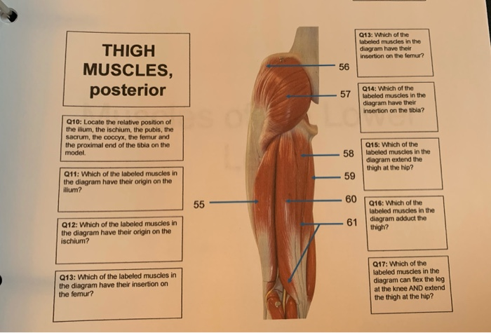
Solved 013 Which Of The Labeled Muscles In The Diagram H Chegg Com from media.cheggcdn.com The piriformis muscle is a key landmark in the gluteal region. The hip muscles cover the hip joint as a muscle sheath. The pubis, ischium, and ilium together constitute the pelvis while the thigh bone is the femur. Muscles are located on opposite sides of the bone it is attached to. This video also provides you with a. If you're just starting your anatomy journey, work on remembering the names of all 11 hip flexor muscles. The hip muscles are all the muscles that act on the hip joint. Muscle and tendon anatomy of the hip (adductors, gluteal muscles (or buttocks), hamstring muscles, femoral cranial nerves (diagrams).
The hip muscles are all the muscles that act on the hip joint.
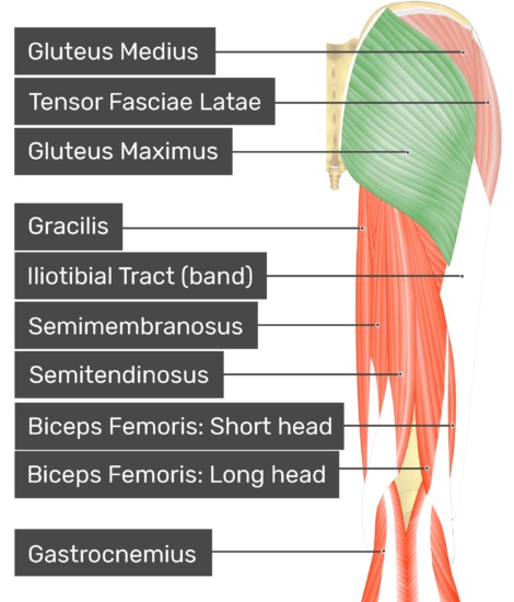
This diagram depicts muscle labeled diagram. Pick which works for you and then we'll review the muscles! Related posts of muscles of the lower back and hip diagram muscle anatomy posterior. Extends from the inner thigh bone to the lumbar vertebrae. Smartdraw includes 1000s of professional healthcare and anatomy chart templates that you can modify and make your own. There are 3 main layers of hip abductor muscles: Muscles are located on opposite sides of the bone it is attached to. Key muscles of the hip : Muscle anatomy guide 12 photos of the muscle anatomy guide anatomy physiology muscle study guide, cat muscle anatomy study guide, human muscle anatomy study guide, muscle anatomy guide, muscle anatomy study guide, human muscles, anatomy physiology muscle study guide, cat muscle anatomy study guide, human. Just need a glimpse, leave your valuable advice let us know , and subscribe us! They also stabilise the hip joint by 'pulling' the femoral head into the acetabulum of the pelvis. Aaa i gg pp r s t. Muscle and tendon anatomy of the hip (adductors, gluteal muscles (or buttocks), hamstring muscles, femoral cranial nerves (diagrams).
This video also provides you with a. The movement that results from contraction is called the action of the muscle. The hip muscles are all the muscles that act on the hip joint. Hip flexion is maximal with a high, forward kick that brings the leg above the level of the waist. Muscular system diagram blank koibanafo.
Gluteus Maximus Attachments Actions Innervation from www.getbodysmart.com Lower back muscle and hip pain may also be caused by stenosis in the spine. Muscle anatomy guide 12 photos of the muscle anatomy guide anatomy physiology muscle study guide, cat muscle anatomy study guide, human muscle anatomy study guide, muscle anatomy guide, muscle anatomy study guide, human muscles, anatomy physiology muscle study guide, cat muscle anatomy study guide, human. The thigh bone or femur and the pelvis join to form the hip joint. The piriformis is the horizontal muscle in the center of the picture running over the top of the sciatic nerve. Typically, one attachment remains stationary and is called the origin and the other attachment moves and is called the insertion. For more anatomy content please follow us and visit. Activity 4.6 labeled muscle diagram. The hip muscles encompass many muscles of the hip and thigh whose main function is to act on the thigh at the hip joint and stabilize the pelvis.without them, walking would be impossible.
The movement that results from contraction is called the action of the muscle.
They can be divided into three main groups: Muscle anatomy guide 12 photos of the muscle anatomy guide anatomy physiology muscle study guide, cat muscle anatomy study guide, human muscle anatomy study guide, muscle anatomy guide, muscle anatomy study guide, human muscles, anatomy physiology muscle study guide, cat muscle anatomy study guide, human. The hip itself is a ball and socket joint, much like the shoulder.the structures necessary to create this joint are the socket, the joint capsule, muscle, ligaments, and the neck. The bones together make up the hip. Scroll down to see the muscle names that go with these letters. We are pleased to provide you with the picture named labelled diagram of the muscles in the human body.we hope this picture labelled diagram of the muscles in the human body can help you study and research. For more anatomy content please follow us and visit our website: Muscles are located on opposite sides of the bone it is attached to. The hip muscles encompass many muscles of the hip and thigh whose main function is to act on the thigh at the hip joint and stabilize the pelvis.without them, walking would be impossible. Muscular system diagram blank koibanafo. If you're just starting your anatomy journey, work on remembering the names of all 11 hip flexor muscles. Pick which works for you and then we'll review the muscles! The majority of muscles in the leg are considered long muscles, in that they stretch great distances.
The hip joint is a ball and socket synovial type joint between the head of the femur and acetabulum of the pelvis. Here are the letters to work with: Smartdraw includes 1000s of professional healthcare and anatomy chart templates that you can modify and make your own. Muscular system diagram blank koibanafo. Most hip pain stems from limited motion of the hip causing abnormal pressures to different muscles, tendons, or ligaments around the area.
Https Www Kean Edu Jeadams Docs Kinesiology Kines Power Points Kines Chap 9 Pdf from They also stabilise the hip joint by 'pulling' the femoral head into the acetabulum of the pelvis. Leg hip muscles diagram / physical therapy guide to groin strain choosept com. Muscles are located on opposite sides of the bone it is attached to. If you're just starting your anatomy journey, work on remembering the names of all 11 hip flexor muscles. The general action of these muscles is to laterally rotate the lower limb. Most hip pain stems from limited motion of the hip causing abnormal pressures to different muscles, tendons, or ligaments around the area. Our latest youtube film is ready to run. This article will introduce the muscles in each group and touch on their origin, insertion, function, and innervation.
The thigh bone or femur and the pelvis join to form the hip joint.
We are pleased to provide you with the picture named labelled diagram of the muscles in the human body.we hope this picture labelled diagram of the muscles in the human body can help you study and research. Functionally, the hip joint enjoys a very high range of motion. Activity 4.6 labeled muscle diagram. Here are the letters to work with: The muscles you probably know the best are your. Lower back muscle and hip pain may also be caused by stenosis in the spine. Muscles are located on opposite sides of the bone it is attached to. Hip muscles diagram the hip muscles cover the hip joint as a muscle sheath. Start studying leg/ hip muscles. Muscles of back of hip an… category: Use acronyms to help you. Related posts of muscles of the lower back and hip diagram muscle anatomy posterior. This article looks at the anatomy of the back, including bones, muscles, and nerves.
There are 3 main layers of hip abductor muscles: hip muscles diagram. The strong muscles of the hip region also help to hold the hip joint together and prevent dislocation.